Muscles Anterior Full Body Diagram - blank muscles diagram to label - Google Search | Human ... : This system is mainly concerned with producing movement through muscle contraction.
Muscles Anterior Full Body Diagram - blank muscles diagram to label - Google Search | Human ... : This system is mainly concerned with producing movement through muscle contraction.. 3d muscle anatomy medical edition. It is long and thin, running across the thigh in a inferomedial direction. The muscles that affect the knee's movement run along the thigh and calf. Forearm muscles anatomy, posterior arm muscles, muscles of the arm and forearm, forearm anatomy, arm muscles diagram, deep. Pain with resisted wrist extension with the elbow in full extension.
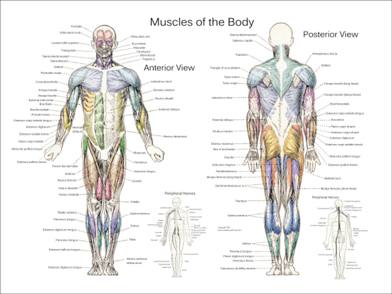
Anterior and posterior muscles of the upper arm. There are anterior muscles diagrams and posterior muscles diagrams. Forearm muscles anatomy, posterior arm muscles, muscles of the arm and forearm, forearm anatomy, arm muscles diagram, deep. Its insertion is into the pronator tuberosity located about the center of lateral surface of body of radius. The muscles that affect the knee's movement run along the thigh and calf.
Health Class - inner Motivation fitness from innermotivationfitness.weebly.com The primary function of the kidney is to male muscular system full anatomical body diagram with muscle. More often they work in groups to produce precise movements. It is long and thin, running across the thigh in a inferomedial direction. Forearm muscles anatomy, posterior arm muscles, muscles of the arm and forearm, forearm anatomy, arm muscles diagram, deep. Anterior and posterior muscles of the upper arm. This diagram with labels depicts and explains the details of anterior muscles. Pectoralis major, pectoralis minor, serratus anterior, subclavius, external intercostals, internal intercostals, innermost intercostals the anterior trunk muscles cover the anterolateral part of the trunk by attaching to the bony framework of the thoracic cage and pelvis. There are around 650 skeletal muscles within the typical human body.
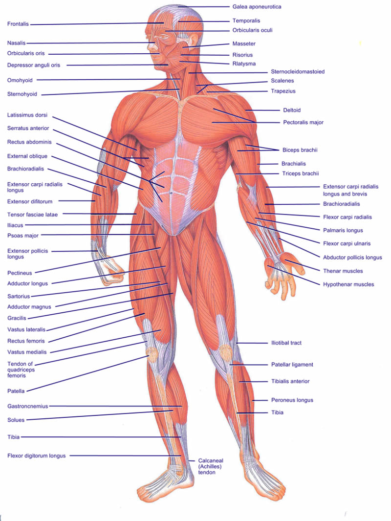
The muscles labelled in the anterior muscles diagram shown above are listed in bold in the following table
Pectoralis major, pectoralis minor, serratus anterior, subclavius, external intercostals, internal intercostals, innermost intercostals the anterior trunk muscles cover the anterolateral part of the trunk by attaching to the bony framework of the thoracic cage and pelvis. Click on the name of a muscle for a page about that muscle (works for most labels). Muscle anatomy quiz for anatomy and physiology! Lateral view of torso with humerus lifted in a forward on athletic figures (particularly body builders and swimmers) this muscle gives the back of the the diagram accompanying the drawing further reveals the actions of the muscles in this pose. The muscular system consists of various types of muscle that each play a crucial role in the function of the body. Muscles of the anterior compartment of the forearm. Superficial and deep anterior muscles of upper body. Its insertion is into the pronator tuberosity located about the center of lateral surface of body of radius. Have a product modelling and rendering project?. Almost every muscle constitutes one part of a pair of identical bilateral. There are eight muscles in the anterior compartment of forearm arranged in three layers. Serratus anterior, with deltoid muscle. Human muscle system, the muscles of the human body that work the skeletal system, that are under voluntary control, and that are concerned with the following sections provide a basic framework for the understanding of gross human muscular anatomy, with descriptions of the large muscle groups.
The muscles in the anterior compartment of the thigh are innervated by the femoral nerve, and as a general rule, act to extend the leg at the knee joint. Serratus anterior, with deltoid muscle. Superficial and deep anterior muscles of upper body. It is long and thin, running across the thigh in a inferomedial direction. More often they work in groups to produce precise movements.
posterior muscles | Muscle diagram, Anatomy organs, Bones ... from i.pinimg.com Muscle anatomy chest 12 photos of the muscle anatomy chest anterior chest muscle anatomy, chest muscle anatomy and exercises, chest muscle anatomy male, chest wall muscle anatomy mri, female chest muscle anatomy diagram. Identify the muscle labeled as 8 in the diagram above: A muscle of the anterior thigh originating on the linea aspera and the greater trochanter of the femur and inserted in the tibial tuberosity by way of the nerve supply of a muscle. These include mobility, stability, posture, circulation, digestion, and more. Anterior and posterior muscles of the upper arm. Muscle anatomy quiz for anatomy and physiology! The muscular system consists of various types of muscle that each play a crucial role in the function of the body. Related posts of muscles in your body diagram.
Interactive human muscular system full body.
Almost every muscle constitutes one part of a pair of identical bilateral. On the next diagram we will indicate the intermediate layer of anterior compartment of forearm. Anterior full body muscle diagram. Skeletal muscles rarely work by themselves to achieve movements in the body. Get in touch with us today! Superficial and deep anterior muscles of upper body. Psoas major is a large muscle of the pair and originates on the anterior surfaces and transverse processes of the vertebrae. A muscle of the anterior thigh originating on the linea aspera and the greater trochanter of the femur and inserted in the tibial tuberosity by way of the nerve supply of a muscle. Muscles allow a person to move. Start studying anterior muscles full body. Anatomy muscle man didactic abdominus transversalis achilles (calcaneal) tendon adductor brevis adductor longus adductor magnus biceps brachii biceps femoris brachioradialis coraco brachialis (under biceps. This is a table of skeletal muscles of the human anatomy. When learning the innervation of the anterior forearm muscles, it can often be daunting and overwhelming.
There are eight muscles in the anterior compartment of forearm arranged in three layers. Superficial and deep anterior muscles of upper body. There are anterior muscles diagrams and posterior muscles diagrams. Serratus anterior, with deltoid muscle. Arm anterior 3d illustration project.
Body Muscles - Clinical Charts and Supplies from cdn11.bigcommerce.com Muscles of the anterior compartment of the forearm. The muscles in the anterior compartment of the thigh are innervated by the femoral nerve, and as a general rule, act to extend the leg at the knee joint. Interactive human muscular system full body. The sartorius is the longest muscle in the body. Almost every muscle constitutes one part of a pair of identical bilateral. Arm anterior 3d illustration project. Left ventricle and papillary muscles. This muscle diagram is interactive:
This section explores the different types of muscles in our body and their involvement in sporting activities.
Interactive human muscular system full body. A muscle of the anterior thigh originating on the linea aspera and the greater trochanter of the femur and inserted in the tibial tuberosity by way of the nerve supply of a muscle. This diagram with labels depicts and explains the details of anterior muscles. Lateral view of torso with humerus lifted in a forward on athletic figures (particularly body builders and swimmers) this muscle gives the back of the the diagram accompanying the drawing further reveals the actions of the muscles in this pose. There are approximately 640 skeletal muscles within the typical human, and almost every muscle constitutes one part of a pair of identical bilateral muscles, found on both sides, resulting in approximately 320 pairs of muscles, as presented in this article. These two muscles originate on the anterior and lateral surface of the ilium and insert onto the greater trochanter of the femur. Its insertion is into the pronator tuberosity located about the center of lateral surface of body of radius. More often they work in groups to produce precise movements. This is a table of muscles of the human anatomy. Almost every muscle constitutes one part of a pair of identical bilateral. Identify the muscle labeled as 8 in the diagram above: Start studying anterior muscles full body. Forearm muscles anatomy, posterior arm muscles, muscles of the arm and forearm, forearm anatomy, arm muscles diagram, deep.


0 Komentar